WHAT an MRI of the Hip Shows
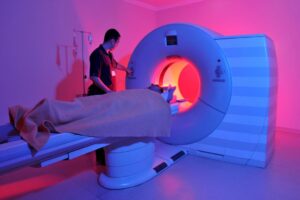
Magnetic resonance imaging is a diagnostic method that provides a detailed picture of the nature of pathological changes in any area of the human body. In orthopedic practice, MRI of the hip joint is most often performed to clarify the causes of coxalgia – pain syndrome in the joint. The method of research does not involve the use of ionizing radiation, which is especially relevant for children and pregnant women. The process of creating clear images, which can be magnified and viewed in volumetric view, is based on magnetic resonance and computer technologies. Contrast enhancement is used to improve imaging capabilities. The diagnostic value of photos obtained after gadolinium injection is comparable to the results of arthroscopy – an invasive study with penetration into the joint cavity with surgical instruments. How is the MRI of the hip joint done? Scans are performed on machines with different magnetic field strengths. The doctor can choose the programs independently depending on the clinical objectives. This is relevant if it is not initially clear what is causing the pain syndrome: pelvic organ pathology or joint pathology. Multiparametric sequences are used in standard situations. Diseases of the hip joint on MRI Magnetic resonance scans are […]
» Read more